
Define Tidal Volume: A Clear Guide for Students and Clinicians
Tidal volume is defined as the volume of air inhaled or exhaled during a single, normal, quiet resting breath, averaging approximately 500 mL in healthy adults. In clinical shorthand, you will see it written as VT. This single measurement sits at the center of respiratory physiology, connecting everything from basic lung function to life-critical decisions in mechanical ventilation. Whether you are a student building your foundational knowledge, an educator designing a lesson on pulmonary physiology, or a respiratory therapist managing a ventilated patient, understanding VT with precision changes how you interpret every breath.
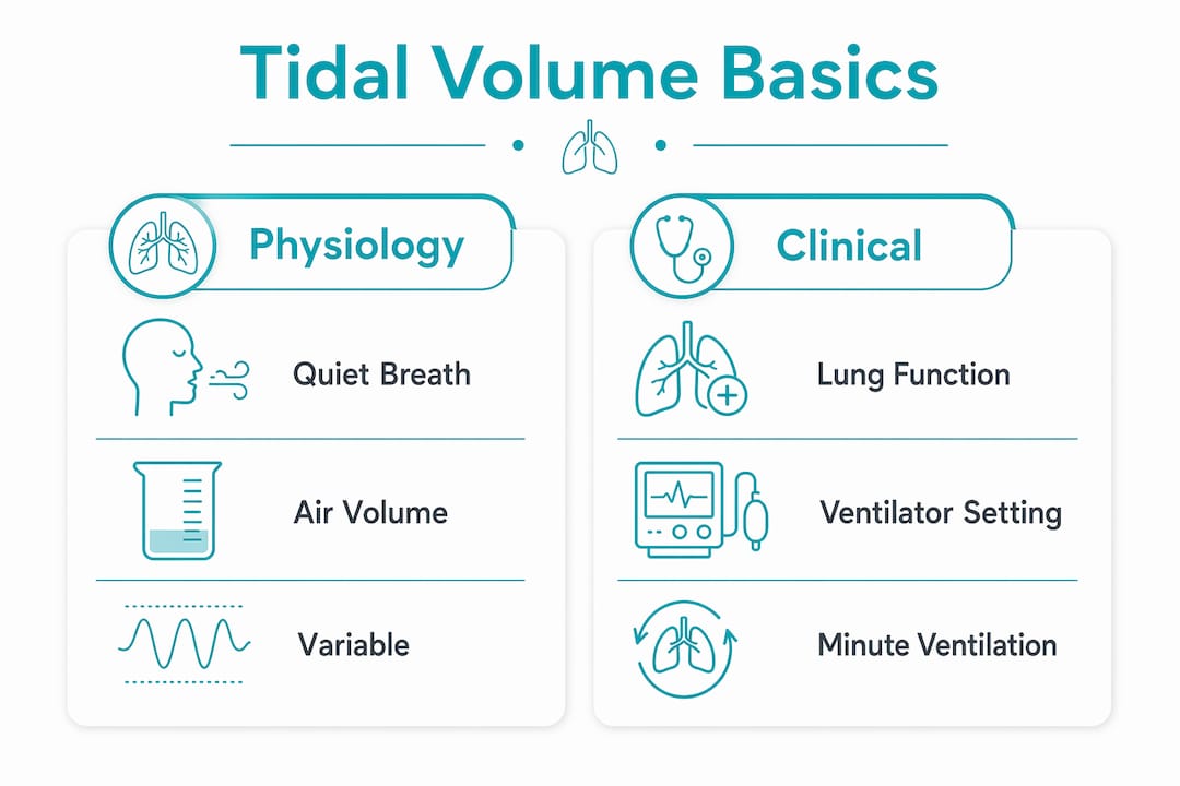
What is tidal volume and how does it fit into lung physiology?
Tidal volume is the most frequently used and most variable lung volume, adapting continuously to physiological conditions rather than staying fixed at a single number. It sits within a hierarchy of lung volumes that together describe the full range of respiratory function. Knowing where VT fits in that hierarchy is the first step toward interpreting any spirometry report or ventilator readout with confidence.
The four primary lung volumes are tidal volume, inspiratory reserve volume (IRV), expiratory reserve volume (ERV), and residual volume (RV). Tidal volume represents only the quiet, effortless portion of breathing. IRV is the additional air you can forcibly inhale above a normal breath, and ERV is the additional air you can forcibly exhale below it. RV is the air that stays in the lungs no matter how hard you exhale.

Two calculations connect tidal volume directly to clinical assessment. Minute ventilation equals tidal volume multiplied by respiratory rate, giving the total air moved per minute. Alveolar ventilation equals tidal volume minus dead space, multiplied by respiratory rate, revealing how much air actually reaches the gas-exchanging alveoli. These two formulas are not interchangeable, and confusing them is one of the most common errors students make.
| Lung Volume or Capacity | Typical Value (Adult Male) | Typical Value (Adult Female) |
|---|---|---|
| Tidal Volume (VT) | ~500 mL | ~400 mL |
| Inspiratory Reserve Volume (IRV) | ~3,000 mL | ~1,900 mL |
| Expiratory Reserve Volume (ERV) | ~1,200 mL | ~800 mL |
| Residual Volume (RV) | ~1,200 mL | ~1,000 mL |
| Vital Capacity (VC) | ~4,700 mL | ~3,100 mL |
| Total Lung Capacity (TLC) | ~5,900 mL | ~4,200 mL |
Pro Tip: When studying lung volumes, memorize the four primary volumes first, then build the capacities by addition. Vital capacity equals IRV plus VT plus ERV. Total lung capacity adds RV to that sum. Building from components prevents confusion on exams and in clinical practice.
How to measure tidal volume in respiratory therapy
Measuring tidal volume accurately in a clinical setting requires more than reading a number off a ventilator screen. The standard bedside method involves collecting total exhaled volume over one minute and dividing by the respiratory rate to calculate average VT. This averaging approach matters because a single breath can be misleading, especially in patients with irregular breathing patterns.
The most critical principle in respiratory therapy is that tidal volume targets use Ideal Body Weight rather than actual body weight. Lung size correlates with height and sex, not with how much a person weighs. Using actual weight in an obese patient inflates the target volume far beyond what the lungs can safely accommodate.
The standard clinical targets follow a clear progression:
- Normal spontaneous breathing: approximately 500 mL or 6 to 8 mL/kg IBW in a healthy adult at rest.
- Standard mechanical ventilation: 6 to 8 mL/kg IBW, balancing adequate gas exchange with lung protection.
- Lung-protective ventilation: 4 to 6 mL/kg IBW, used in conditions such as Acute Respiratory Distress Syndrome (ARDS) where alveoli are fragile and prone to overdistension.
- Monitoring for VILI: Ventilator-induced lung injury (VILI) develops when volumes exceed safe limits, causing barotrauma and volutrauma to already-compromised lung tissue.
Ventilator-induced lung injury risk rises sharply when clinicians set volumes based on actual weight in larger patients. A 120 kg patient with a height-based IBW of 70 kg could receive nearly double the safe tidal volume if actual weight drives the calculation. That error is not theoretical. It has measurable consequences for patient outcomes.
Pro Tip: Always calculate IBW before setting any ventilator tidal volume. For males, IBW in kg equals 50 plus 2.3 multiplied by each inch over 60 inches of height. For females, start at 45.5 instead of 50. This calculation takes under 30 seconds and removes one of the most preventable sources of ventilator harm.
Why tidal volume is not a fixed number
One of the most important things to understand about tidal volume is that it changes constantly. Tidal volume fluctuates significantly with activities such as talking, exertion, anxiety, and sleep, which means clinicians interpret averages over time rather than isolated single-breath measurements. A student who memorizes 500 mL and treats it as a constant will misread clinical data every time.
Consider these real shifts in VT across common physiological states:
- Exercise: VT can rise from 500 mL to over 2,000 mL in vigorous activity, working in combination with increased respiratory rate to multiply minute ventilation several times over.
- Sleep: VT typically decreases slightly during non-REM sleep as metabolic demand falls.
- Anxiety or pain: Both increase respiratory rate and can alter VT, sometimes producing rapid shallow breathing that compromises gas exchange.
- Talking: Even speaking aloud modifies the breathing pattern, interrupting the normal rhythm and temporarily reducing effective VT.
The role of anatomical dead space makes this variability even more clinically significant. Approximately one-third of tidal volume remains in the conducting airways, including the trachea, bronchi, and bronchioles, and never reaches the alveoli where gas exchange occurs. With a typical VT of 500 mL, roughly 150 mL is dead space volume. Only 350 mL participates in actual gas exchange.
“Recognizing that approximately 30% of tidal volume remains in dead space is essential to understanding why shallow, rapid breaths can cause hypoventilation despite seemingly normal tidal volume numbers.” — Respiratory Physiology, Dead Space Volume
This is why a patient breathing at 20 breaths per minute with a VT of 300 mL may appear to have adequate minute ventilation on paper, yet still be hypoventilating at the alveolar level. The math only tells the full story when dead space is subtracted.
How does tidal volume compare to vital capacity and other lung volumes?
Tidal volume and vital capacity are frequently confused by students, yet they describe completely different aspects of lung function. Tidal volume is the air moved during quiet breathing, while vital capacity is the maximum volume of air a person can exhale after a maximum inhalation. Vital capacity represents the upper boundary of voluntary respiratory effort. Tidal volume represents the baseline of effortless breathing.
A common misconception is that a large tidal volume indicates strong lungs. In reality, a high VT during quiet breathing can signal respiratory distress, where the body is working harder than normal just to maintain baseline ventilation. Vital capacity is the better indicator of overall lung reserve and respiratory muscle strength.
| Measurement | What It Represents | Typical Adult Value |
|---|---|---|
| Tidal Volume (VT) | Air per quiet breath | ~500 mL |
| Vital Capacity (VC) | Max exhale after max inhale | ~4,700 mL (male) |
| Inspiratory Capacity (IC) | VT plus IRV | ~3,500 mL (male) |
| Functional Residual Capacity (FRC) | ERV plus RV | ~2,400 mL (male) |
| Residual Volume (RV) | Air remaining after full exhale | ~1,200 mL (male) |
The clinical relevance of each measurement differs by context. VT guides ventilator settings and monitors breathing effort. Vital capacity tracks neuromuscular disease progression in conditions like Guillain-Barré syndrome. Functional residual capacity reflects the resting lung position and is altered in obesity and ARDS. Treating these values as interchangeable leads to misinterpretation.
Pro Tip: When reviewing a pulmonary function test, locate VT and vital capacity separately and compare them as a ratio. If VT is consuming an unusually large proportion of vital capacity, the patient has limited respiratory reserve, which is a clinically significant finding regardless of whether VT itself looks normal.
Key takeaways
Tidal volume is a dynamic, context-dependent measurement that drives both basic respiratory physiology and critical clinical decisions, particularly in mechanical ventilation using Ideal Body Weight as the calculation standard.
| Point | Details |
|---|---|
| Core definition | Tidal volume is the air volume per quiet breath, averaging 500 mL in healthy adults. |
| Dead space impact | Roughly 150 mL of each breath stays in conducting airways and never reaches alveoli. |
| IBW-based calculation | Clinical tidal volume targets use Ideal Body Weight, not actual weight, to prevent lung injury. |
| Lung-protective target | ARDS and fragile lung conditions require 4 to 6 mL/kg IBW to reduce overdistension risk. |
| VT vs. vital capacity | Tidal volume reflects quiet breathing effort; vital capacity reflects maximum respiratory reserve. |
Why I think students underestimate the dead space problem
When I look at how tidal volume is typically taught, the 500 mL figure gets all the attention, and dead space gets a footnote. That imbalance creates a real gap in clinical reasoning. Students who focus only on tidal volume numbers without accounting for dead space consistently overestimate how much ventilation is actually reaching the alveoli, and that mistake follows them into practice.
The most useful shift I have seen in learners is when they stop thinking of tidal volume as a target to hit and start thinking of it as one variable in a system. Alveolar ventilation is the outcome that matters. Tidal volume is just one input. Respiratory rate, dead space, lung compliance, and patient position all shape the final result.
My honest advice for students: practice the alveolar ventilation calculation until it is automatic. Take any tidal volume, subtract 150 mL for dead space, multiply by respiratory rate, and ask whether that number meets the patient’s metabolic demand. That habit, more than any memorized reference value, is what separates a competent clinician from a great one.
For educators, the most powerful teaching moment is showing students a patient scenario where minute ventilation looks normal but alveolar ventilation is critically low. That contrast makes the dead space concept unforgettable in a way that no textbook definition can replicate.
— Tita
Bring science concepts to life with hands-on discovery
Understanding respiratory physiology starts with curiosity, and curiosity grows fastest when learning becomes an experience rather than a lecture. Teamgeniussquad builds that kind of learning through hands-on, screen-free STEAM discovery kits designed to help young learners step into the role of scientists and innovators.

Whether you are an educator looking for tools that make science tangible or a parent wanting to spark a child’s love of discovery, Teamgeniussquad’s experiment kits bring concepts to life through real, purposeful play. The STEM-STEAM Electricity Lab Bundle is a standout example of how structured, hands-on exploration builds both knowledge and confidence. Powered by the proprietary E³ Method (Engage, Encourage, Empower), every kit is designed to help children see themselves as capable, creative problem-solvers from the very first experiment.
FAQ
What is the normal tidal volume for a healthy adult?
The normal tidal volume for a healthy adult is approximately 500 mL per breath during quiet, resting respiration. Adult males average around 500 mL, while adult females typically range from 400 to 500 mL.
How is tidal volume calculated in mechanical ventilation?
Tidal volume in mechanical ventilation is calculated using Ideal Body Weight rather than actual body weight, with standard targets of 6 to 8 mL/kg IBW. Lung-protective ventilation protocols, used in conditions like ARDS, target 4 to 6 mL/kg IBW to reduce the risk of ventilator-induced lung injury.
What is the difference between tidal volume and minute ventilation?
Tidal volume is the air moved in a single breath, while minute ventilation is tidal volume multiplied by the respiratory rate, representing total air moved per minute. The two values are related but serve different clinical purposes, with minute ventilation reflecting overall breathing workload.
Why does dead space matter when interpreting tidal volume?
Approximately 150 mL of each tidal volume breath remains in the anatomical dead space of the conducting airways and does not participate in gas exchange. This means effective alveolar ventilation is always less than the measured tidal volume, which is why shallow rapid breathing can cause hypoventilation even when total ventilation appears adequate.
How does tidal volume differ from vital capacity?
Tidal volume is the air moved during normal, effortless breathing, averaging around 500 mL. Vital capacity is the maximum volume of air exhaled after a full inhalation, averaging around 4,700 mL in adult males, and it reflects the total usable lung reserve rather than resting breathing effort.


Leave a comment
This site is protected by hCaptcha and the hCaptcha Privacy Policy and Terms of Service apply.